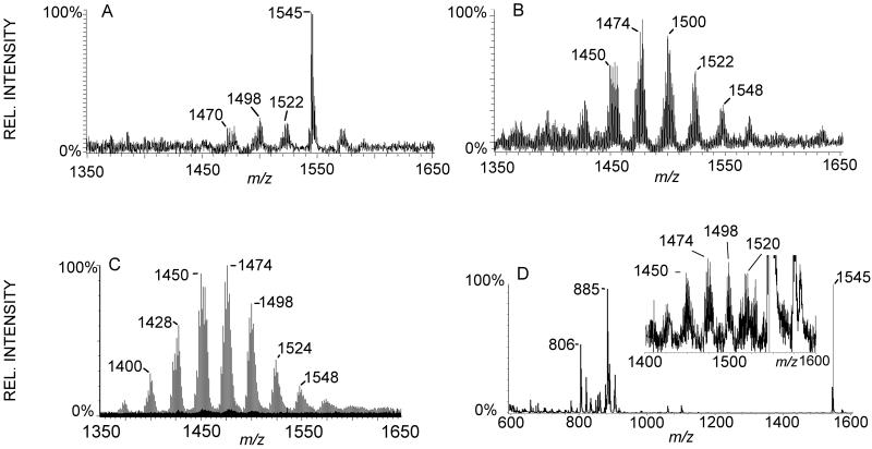
Fig 2.
Comparison MALDI-MS and ESI-LC-MS from pnd 17 rat brain. A: MALDI-MS of brain total lipid extract barely detects some CL species predominantly as [M-2H+Na]− adducts. Ganglioside GM1 at m/z 1545 obscures CL in this region. B: Split-flow LC-MS allowed collection of this CL fraction and subsequent analysis by MALDI-MS resulting in more species of CL detected, and these were mixtures of [M-H]− and [M-2H+Na]− adducts. C: ESI-LC-MS of same total lipid extract detects 70 species of CL, and these were detected as [M-H]− ions. D. On—tissue phospholipase C (PLC) treatment and subsequent washing results in MALDI-MSI detection of several species of CL predominantly as [M-H]− ions. The inset region is at 50× magnification. MALDI-MS and –MSI were acquired with an Autoflex in negative mode and ESI-LC-MS with a Q-TOF in negative mode.

