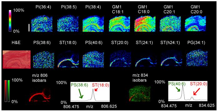
Fig. 5.
FT-ICR MSI completely resolves isobaric rat brain species. Several lipid species from various classes were detected at high mass resolution and their localizations correlated with an H&E stain of the tissue section. The white bar is 1000 microns and the scale displays relative intensities with respect to the abundance of each ion. Two sets of isobaric species (m/z 806.5 and 834.5) were completely resolved. The ions at m/z 806.497 and m/z 806.545 representing PS(38:6) and ST(d18:1/18:0) were resolved from each other, as were the ions at, m/z 834.528 and m/z 834.576 representing PS(40::6) and ST(d18:1/20:0), respectively. Each of these isobaric pairs was overlaid on the same MALDI image, but at the same absolute scale. Green pixels represented the PS species, and red represented the ST species. This confirmed that the m/z 806 species consisted almost entirely of ST(d18:1/18:0), while the m/z 834 species was mostly PS(18:0/22:6) but with a significant amount of ST(d18:1/20:0). The summation MALDI-MSI spectrum from the entire image further confirmed these results. MSI images were acquired with a Solarix in negative mode.

