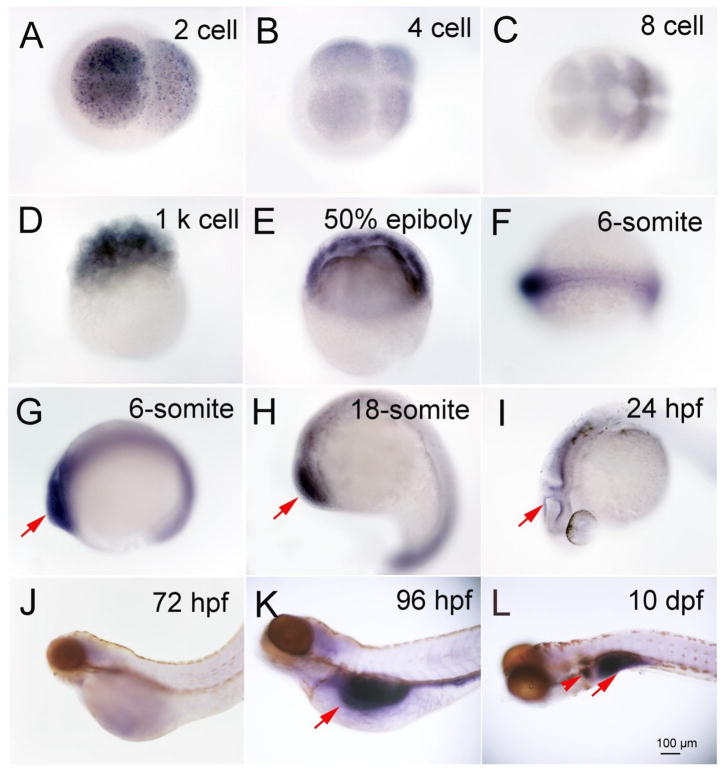
Fig. 1.
Expression of Deiodinase 1 in different developmental stages of zebrafish embryo and larva as shown by dark coloration using in situ hybridization. A, 2 cell; B, 4 cell; C, 8 cell; D,1 k cell; E, 6-somite; F and G, 6-somite; H, 18-somite; I, 24 hpf; J, 72 hpf; K, 96 hpf; L, 10 dpf. Red arrows indicate the location of expression.

