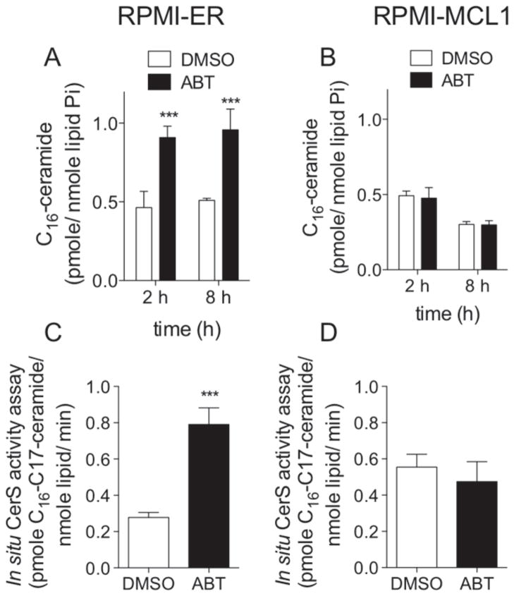
Figure 3. MCL1 inhibits ABT-263 induced C16-ceramide generation and increased in situ CerS activation in RPMI8226 human plasma cell leukemia.
A, RPMI8226 cells stably expressing GFP and a portion of an irrelevant protein, ER. (RPMI-ER) were treated with 0.5 μM of ABT-263, or vehicle (DMSO), for 2 and 8 hours and C16-ceramide quantified by HPLC-MS/MS. B, RPMI8226 stably expressing GFP and MCL1 (RPMI-MCL1) were treated with 5 μM of ABT-263, or vehicle (DMSO), for 2 and 8 hours and C16-ceramide quantified by HPLC-MS/MS. C. RPMI-ER cells were incubated with 0.5 μM of ABT-263, or vehicle (DMSO), for 1 hour and 45 minutes. 1μM C17-sphingosine was then added to the media for an additional 15 minutes. Cells were immediately lysed and the amount of C17-ceramide in the cells were quantitated by HPLC-MS/MS. D, RPMI-MCL1 cells were incubated with 5 μM of ABT-263, or vehicle (DMSO), for 1 hour and 45 minutes. 1μM of C17-sphingosine was then added to the media for an additional 15 minutes. Cells were immediately lysed and the amount of C17-ceramide in the cells were quantitated by HPLC-MS/MS. Lipids were normalized to total lipid phosphate and data expressed as a fold-change of the untreated control. All data points represent the compilation of at least biological triplicates. ***, p=<0.0005; **, p=<0.005; *, p=<0.05.

