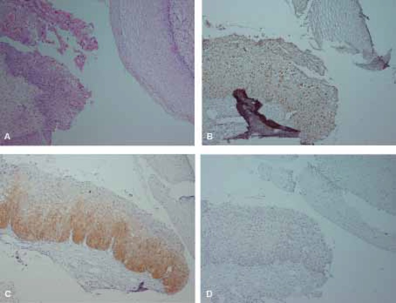
Figure 1.
Hematoxylin and Eosin (H&E) and immunohistochemical staining of Ki67, p16 and CK17 in CIN1. A, H&E staining. B, scattered Ki67 immunostaining in CIN1 and negative in normal epithelium. C, diffuse (one-third) p16 immunostaining in CIN1 and negative staining in normal epithelium. D, negative staining for CK17 in both normal tissue and CIN1. (×100)

