Figure 1.

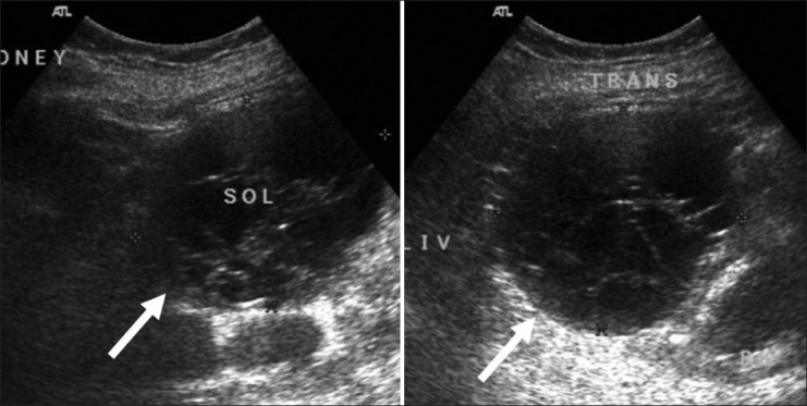
Ultrasound of the right kidney. Longitudinal (left) and transverse (right) images show a well-defined multiseptated cystic lesion (arrows) at the lower pole

Ultrasound of the right kidney. Longitudinal (left) and transverse (right) images show a well-defined multiseptated cystic lesion (arrows) at the lower pole