
Figure 1.
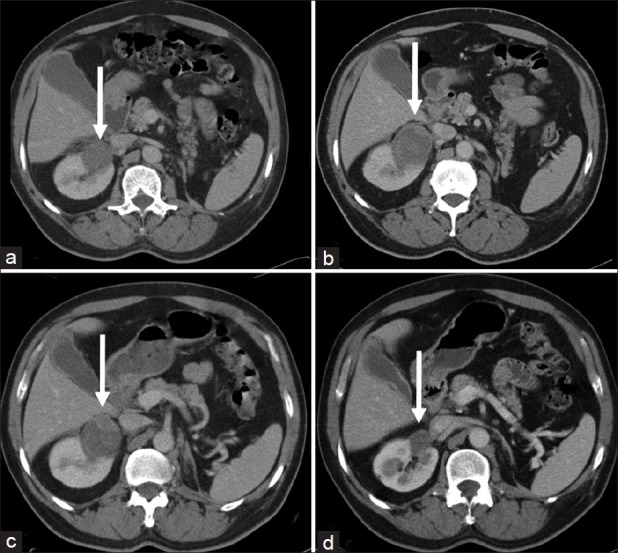
Contrast-enhanced CT scan depicting renal mass or ablation site (white arrow). (a) Pre-op 4.3-cm renal mass. (b) MWA post-op scan showing ablated tumor. (c) MWA post-op film showing recurrence at anterior portion of ablation zone. (d) RFA post-op scan shows no residual disease at 24 months