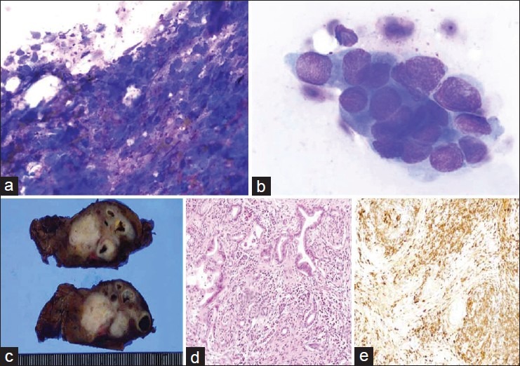
Figure 1.

(a) Numerous lymphocytes and plasma cells with necrotic material and fibrosis are visible (Diff-Quik, ×400); (b) Atypical epithelial cells (Diff-Quik, ×1000); (c) The cut surface of the resected specimen show an elastic, hard, white mass that was located in the pancreas tail; (d) Ductal adenocarcinoma with lymphoplasmacytic infiltration was visible (H and E, ×100); (e) Immunohistochemical findings showed abundant IgG4-positive plasma cells around the pancreatic duct and cancer cells (IHC, ×100)
