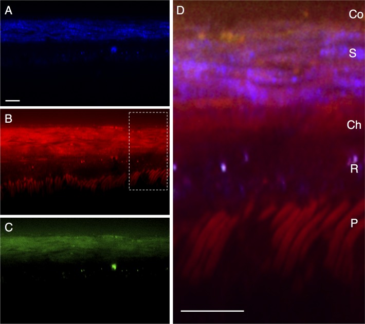
Figure 2.
Line scan of a posterior region of a mouse eye, representing a transverse/sagittal section. The conjunctiva (Co) is located at the top of the images, with retinal photoreceptors (P) located at the bottom. (A) shows two-photon autofluorescence (TPAF) in blue. The coherent anti-Stokes Raman scattering (CARS) signal from the forward detector (F-CARS, red) is shown in (B) and from the epi-detector (E-CARS, green) in (C). Both CARS and TPAF signals are overlapped in (D). S, sclera; Ch, choroid; R, RPE; P, photoreceptors. Scale bar: 10 μm.

