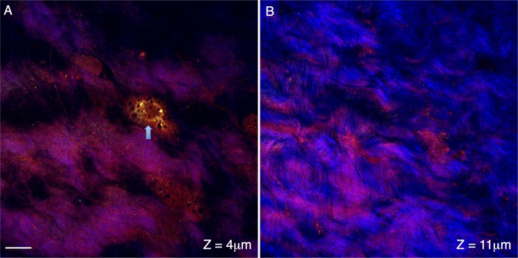
Figure 3.
Cross-sections of conjunctival and sclera regions of the posterior mouse eye. Signal from the forward coherent anti-Stokes Raman scattering detector (F-CARS, red), epi-detector (E-CARS, green) and two-photon autofluorescence (TPAF, blue) are shown. The cross-section in (A) shows the surface region of the mouse eye ∼4 μm from the surface of the tissue. A surface cell is apparent in the CARS channels (arrow) and is likely a bulbar conjunctival cell but may be a scleral fibroblast. The cross-section in (B) travels through the mouse sclera (∼11 μm from the tissue surface). The collagen fibers appear in the TPAF channel as repeated strands of material interwoven into one another. Scale bar: 10 μm.

