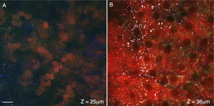
Figure 4.
Cross-sections of the choroid and retinal pigment epithelium. Signal from the forward coherent anti-Stokes Raman scattering detector (F-CARS, red), epi-detector (E-CARS, green), and two-photon autofluorescence (TPAF, blue) are shown. A cross-section of choroid is shown in (A), located approximately 25 μm from the tissue surface. The spherical objects with F-CARS signal are likely red blood cells within the choriocapillaris. The cross-section in (B) travels through a RPE, approximately 36 μm from the surface of the tissue. Dark regions lacking CARS/TPEF signal are of a size and spacing to be nuclei. Punctate objects (∼1 μm) with strong CARS/TPEF signal (white due to the chromatic combination of the red/green/blue channels) may represent retinyl ester storage structures. Scale bar: 10 μm.

