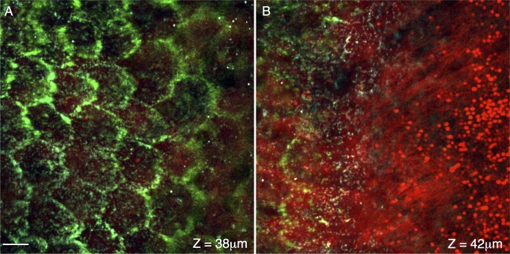
Figure 5.
Cross-sections at the boundary of the retinal pigment epithelium and photoreceptors. Signal from the forward coherent anti-Stokes Raman scattering detector (F-CARS, red), epi-detector (E-CARS, green), and TPAF (blue) are shown. (A, B) are located approximately 38 and 42 μm from the surface of the eye. The tight-junctions of the RPE, with their characteristic hexagonal shape, are visible in the E-CARS channel (green, [A]). (B) shows the transition of the RPE tight junctions (left, green) to the lipid-rich RPE microvilli (center, red) to the distal ends of the photoreceptor outer segments (red, right). Scale bar: 10 μm.

