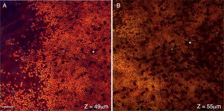
Figure 6.
Cross-sections of the distal ends of the photoreceptor layer. Signal from the F-CARS (red), E-CARS (green), and TPAF (blue) are shown. Cross-sections shown in (A, B) are located approximately 49 and 55 μm interior to the surface of the eye, travel through the distal and middle regions of the photoreceptor outer segments, respectively. These lipid rich disks generate a strong F- but not E-CARS signal. Occasional dark spaces within the outer segments (*) likely represent the location of cone photoreceptors. Scale bar: 10 μm.

