Figure 2.
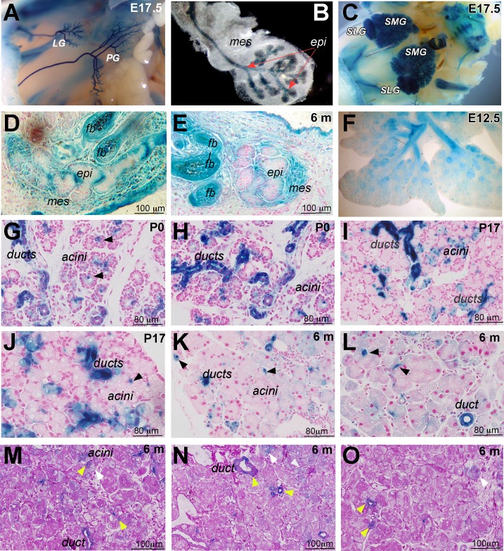
Analysis of Runx1 expression in the lacrimal gland of heterozygous Runx1 knockin-lacZ mice. (A, B) Whole-mount X-Gal staining of mouse embryo at E17.0. Runx1 expression is restricted to the epithelium of the LG and PG; (B) shows an excised LG. Red arrows mark epithelial component of the lacrimal gland. (C) Runx1-lacZ is expressed in the SLG and SMG. (D, E) Runx1 is also expressed in the epithelial and mesenchymal components of the meibomian gland (mei) and lung epithelium (F). (G–L) Expression pattern of Runx1 in the LGs at postnatal development day 0 (P0; [G, H]), P17 (I, J), 6-month-old (K, L) old mice. Runx1 expression is restricted to the ducts and centroacinar cells (black arrows) within the secretory acini. LGs obtained from Runx1 knockin-lacZ mice were stained with X-Gal and paraffin sections were prepared. Nuclei were stained with fast red to visualize LG morphology. (M–O) PAS reaction was also used for staining of sections of Runx1-LacZ LGs. Strong LacZ staining was seen within all PAS+ ducts, including a main duct (m. duct) and centroacinar cells (yellow arrows) and within some PAS– acini/cells (white arrows).

