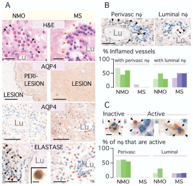
FIGURE 5.
Abundant neutrophil elastase within human NMO but not MS lesions. (A) NMO vs MS brain lesions. H&E: Arrows = neutrophils (black), eosinophils (blue), mononuclear cells (orange). AQP4 immunostain: (Top) Lesion and perilesion, (Bottom) Vessel in lesion. Lu = lumen. NE immunostain: Perivascular region. Arrows = NE+ (black) and NE− (orange) leukocytes. (Inset) NE+ cell. (B) (Top) Vessel with perivascular (no intraluminal) and 1 with intraluminal (no perivascular) neutrophils. (Bottom) % inflamed vessels with perivascular NE+ cells and % with circulating (luminal) NE+ cells in 3 NMO (green) vs 3 MS (purple) samples. (C) Top (i) Inert and (ii,iii) active neutrophils. (i) NE (black arrows) is intracellular (red arrows). (ii) Extracellular NE. (iii) Elongated neutrophil (black arrows). (Bottom) Number of perivascular and luminal active neutrophils in 3 NMO (green) and 3 MS (purple) samples. Bar = 20μm (A: H&E), 0.5mm (A: AQP4 top), 100μm (A: AQP4 bottom), 50μm (A: Elastase, B), 5μm (A: Elastase inset, C).

