Figure 6.
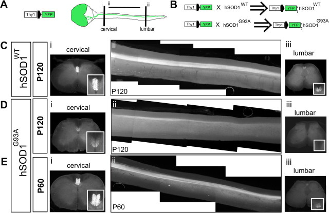
The corticospinal tract degenerates in Thy1-YFP;hSOD1G93A mice. A, Breeding of hSOD1 transgenic mice with Thy1-YFP transgenic mice was used to visualize the CST in hSOD1G93A and hSOD1WT mice. Schematic drawing of cerebral cortex and spinal cord of Thy1-YFP transgenic mice, in which the CST is visualized by YFP fluorescence in the spinal cord. Cervical and lumbar segments of the spinal cord were investigated by single 50-μm-thick axial sections (i, iii). Sagittal sections were used for the thoracic cord; to identify the entire extent of the CST in the thoracic segment, all 150-μm-thick sagittal sections containing CST axons were digitally collapsed into a single two-dimensional image (ii). B, Thy1-YFP;hSOD1G93A and Thy1-YFP;hSOD1WT mice (hemizygous for both the Thy1-YFP and the hSOD1G93A genes) were generated by serial breeding Thy1-YFP mice and hSOD1G93A or hSOD1WT mice, respectively. Thy1-YFP;WT mice were used as controls. C–E, Insets (Ci, Di, Ei, Ciii, Diii, Eiii) show higher-magnification views of the dorsal funiculi, indicating CST axons at both the cervical (i) and lumbar (iii) segments of the spinal cord. C, At P120, the CST did not show any sign of degeneration in Thy1-YFP;hSOD1WT mice. The CST is normal in the cervical (i), thoracic (ii), and lumbar spinal cord (iii). D, E, In Thy1-YFP;hSOD1G93A mice, CST degeneration is most dramatic between P60 and P120. There is substantial loss of cervical (Di) and thoracic (Dii) CST axons by P120, and innervation of the lumbar spinal cord is drastically reduced to absent in Thy1-YFP; hSOD1G93A mice at P120 (Diii).

