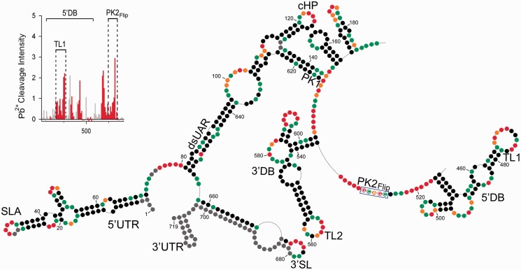
Figure 4.
Chemical probing of PK2Flip mutant RNA. The description of the scheme follows the convention of the Figure 1 legend. The flipping mutation within PK2 is marked by a blue rectangle. The insert in the left top corner represents increased Pb2+-induced hydrolysis within PK2 and TL1 regions.

