
Figure 1.
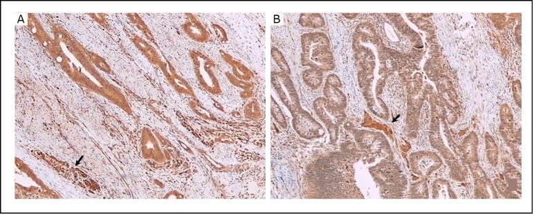
LC3B protein was detected in human colorectal cancer tissues by immunohistochemical staining with LC3B antibody. A: LC3B expression in cancer cells showing equal or stronger intensity than in nerve cells (arrow) were deemed “strongly positive.” B: LC3B expression in cancer cells showing weaker intensity than in nerve cells (arrow) were deemed “weakly positive”. Original magnification, × 40.