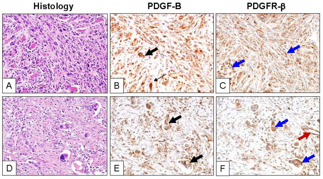
Figure 1.
Representative microphotographs of two sarcomatoid lung carcinomas (A, pleomorphic subtype, and D, giant cell, hematoxilyn-eosin, H&E) with strong cytoplasmic expression of PDGF-B (B and E; black arrows) and PDGFR-β (C and F; black arrows) in highly atypical malignant cells. Tumor stromal cells and blood vessel endothelial cells show PDGFR-β IHC expression (red arrow). Magnification of ×200.

