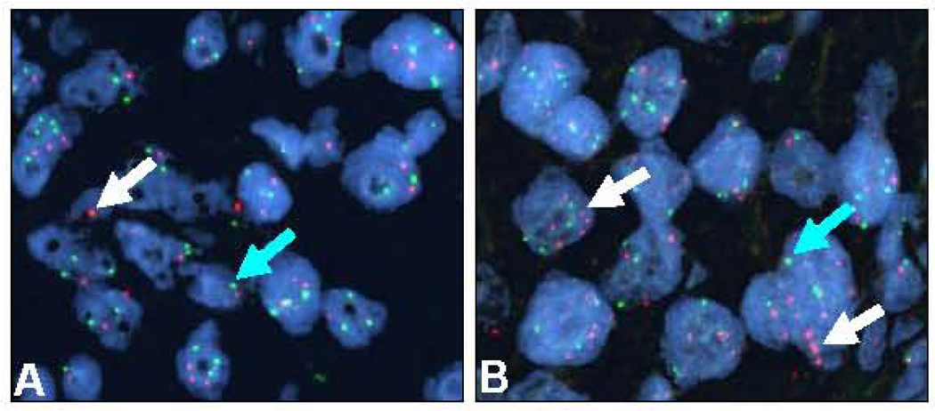
Figure 3.
Representative microphotographs of two sarcomatoid lung carcinomas examined by FISH showing normal PDGFRB copies (A) and gene amplification (B) in malignant cells. Magnification ×1,000. Red signals (white arrows) represent PDGFRB gene copies and green signals (green arrows) represent the chromosome 5 centromeric probe. Cell nuclei stained with 4’,6-diamidino-2-phenyldole.

