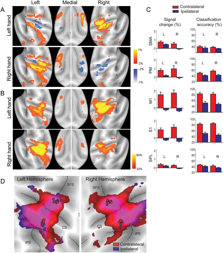
Figure 2.
Representation of contra- and ipsilateral finger presses in the human neocortex. (A) Group-average percent signal change (threshold ±0.2%) averaged over all fingers and compared with rest. During ipsilateral actions, suppression can be observed in primary sensory and motor cortices. Positive activation during ipsilateral finger presses can be observed in the left hemisphere. (B) Classification accuracy, thresholded at >32%, Z > 1.97. Colored regions show local voxel patterns that significantly distinguish between different fingers. High classification accuracy for ipsilateral presses can be found in regions that are deactivated compared with the rest. (C) Mean signal change and classification accuracy for contralateral (red) and ipsilateral (blue) finger presses in the informative region within 5 anatomically defined ROIs of the left (L) and right hemispheres (R). Error bars indicate across-subject SE. (D) Overlap of classification accuracy (>32%) for contralateral (red) and ipsilateral (blue) fingers. Circles indicate the COG of classification accuracy for individual participants for precentral and postcentral ROIs. CS, central sulcus; SFS, superior frontal sulcus.

