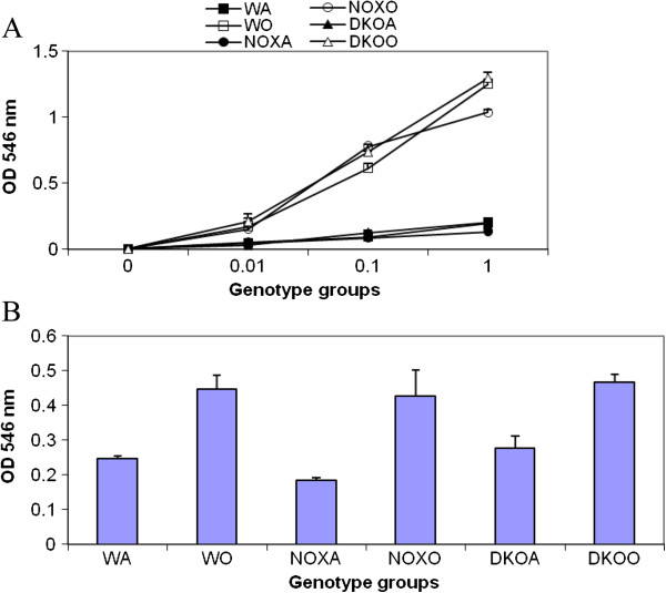
Figure 4.
T cells response in WT vs. KO mice. A. Splenocytes from control (saline treated) and OVA treated mice were made into single cell suspensions in DMEM+10% heat-inactivated FCS. 0.1 million cells were plated per well without and with increasing concentrations of anti-CD3 antibody and a constant concentration of anti-CD28 antibody (1μg/ml) and cultured for 3 days. B. 1μM PMA and 10ng/ml ionomycin was used to stimulate splenocytes from the above experimental mice and proliferation measured after 3 days. To measure proliferation, MTT assay called CellTiter96 (Promega) was used. OD 546 nm is directly proportional to the number of cells in culture. Abbreviations used are: WT=wildtype, NOX=gp91phox−/−, DKO=gp91phox-MMP-12 double knockout, WA=WT+alum, WO=WT+OVA, NOXA=gp91phox−/−+alum, NOXO=gp91phox−/−+OVA, DKOA= gp91phox-MMP-12 double knockout+alum, DKOO= gp91phox-MMP-12 double knockout+OVA. Data presented are average of 3 independent experiments ± SEM. (n=5/group).

