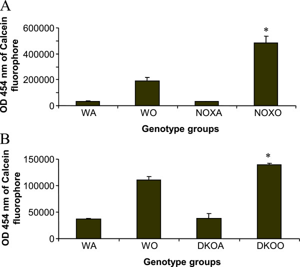
Figure 6.
Inhibition of MCP-1-driven chemotaxis of alveolar macrophages in post-OVA KO mice. 15mM MCP-1 was put in 29μl volume in the lower well and 10 × 106 alveolar macrophages (from 4 mice/experimental group), also in 29μl volume in the upper wells of a 96 well Neuroprobe CTX plates (Chemicon) in high glucose medium for 2 h followed by detachment by mechanical scraping and resuspension in Phenol red-free high glucose DMEM (Gibco) with 5% FBS with 0.5μg/ml Calcein-AM (1:2000 dilution) and incubation for 20 min at 370C. Migrated cells were quantified by fluorescence (excitation at 488 nm, emission at 520 nm) using a Victor 3V (Perkin Elmer laboratories) using a Wallac1420 software. 2.5-fold and 1.26-fold increase in OD (proportinate to number of fluorescing cells in the upper well equivalent to the number of cells migrated) was found in post-OVA gp91phox−/− and DKO mice respectively. * denotes p value<0.05 compared to values in OVA-treated wildtype group.

