
FIGURE. IMAGING AND PATHOLOGICAL FEATURES OF CORTICOBASAL DEGENERATION.
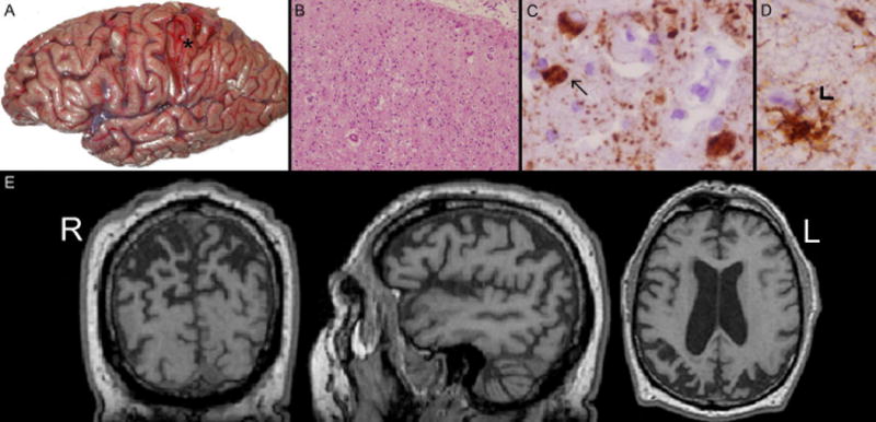
Panel A: Gross atrophy in parietal lobe (asterisk) of Case 1; Panel B: H & E preparation of the angular gyrus of Case 1 at 10X magnification showing significant neuronal dropout and gliosis in the superficial cortical layers; Panel C: PHF stain for tau in the cortex of the angular gyrus of Case 1 at 60X magnification showing balloon cells (arrow); Panel D: PHF stain for tau in the white matter of the angular gyrus of Case 1 at 60X magnification showing an astrocytic plaque (arrowhead); Panel E: T1 MPRAGE MRI sequence showing bilateral parietal-occipital atrophy in coronal (y = −60), sagittal (left hemisphere) (x = −47), and axial (z = +14) views of Case 2.