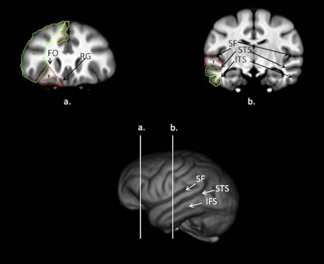
Figure 1.
Bottom, 3D-reconstruction of chimpanzee template brain with vertical white lines representing regions outlined above (a, b). a, Coronal view of the chimpanzee brain with the orbital (red), dorsal (green), and mesial (yellow) prefrontal cortex regions outlined on the scan. RG, Rectus. b, Coronal view of the chimpanzee brain with the superior (red), middle (green) and inferior (yellow) temporal gyri regions outlined on the scan.

