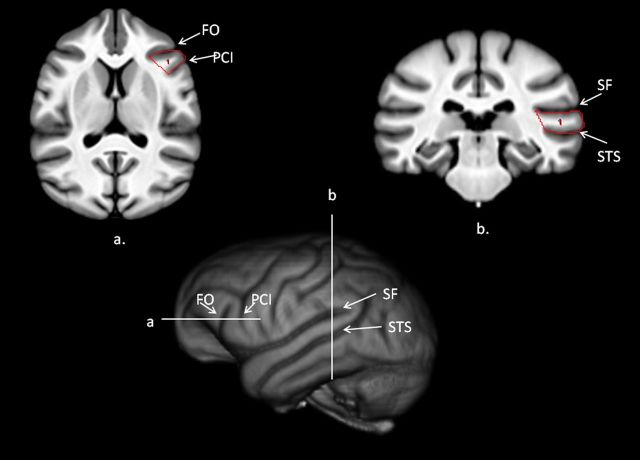
Figure 3.
Bottom, 3D-reconstruction of chimpanzee template brain with horizontal and vertical white lines representing regions outlined above (a, b). a, Transverse view of the chimpanzee template brain with the IFG outlined on the scan. b, Coronal view of the chimpanzee template brain with the posterior superior temporal gyrus outlined on the scan.

