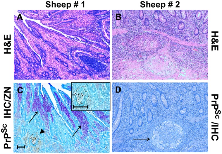
Figure 3. Inflammatory lesion patterns and PrPSc deposition in the ileum of sheep with scrapie and paratuberculosis.
Sheep #1: Granulomatous enteritis is shown, with inflammatory gut lesions exhibiting a “lepromatous” morphology (A); by means of IHC, PrPSc deposition is apparent inside a constitutive lymphoid follicle of ileal Peyer’s patches (arrowhead), while no PrPSc-positive immunostaining is detectable within the surrounding granulomatous inflammatory foci, in which consistent numbers of acid-fast bacilli (M. avium subsp. paratuberculosis, MAP) are present inside epithelioid macrophages (arrows); a higher magnification of the above lymphoid follicle, harboring PrPSc aggregates, is shown in the inset (C). Sheep # 2: Granulomatous enteritis is shown, with inflammatory gut lesions exhibiting a “tuberculoid” morphology (B); no IHC evidence of PrPSc deposition is observed inside a microgranuloma (D, arrow). H&E stain (A-B); Ziehl-Neelsen stain and PrPSc IHC with F99 as primary antibody (C, see also “Materials and Methods”); PrPSc IHC with F99 as primary antibody and Mayer’s hematoxylin counterstain (D). Scale bars = 100 µm and 50 µm (inset of C).

