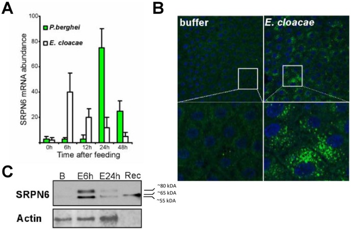
Figure 3. Activation of SRPN6 midgut expression by E. cloacae.
(A) Time course of An. stephensi SRPN6 mRNA expression after feeding of E. cloacae (1×10⁶/ml; open bars) or P. berghei (green bars) as determined by qRT-PCR using ribosomal protein S7 (rpS7) for normalization. Values are reported in fold change relative to expression before feeding (0 h). (B) Immunolocalization of SRPN6 in midguts of mosquitoes fed with buffer (left panels) or E. cloacae (right panels). Guts were dissected 6 h after feeding, opened up into sheets, fixed and the protein detected with an anti-SRPN6 antibody. The inserts show higher magnifications of the areas within the squares. (C) Western blot analysis of SRPN6 protein expression after feeding with buffer alone (B) or at 6 and 24 h after E. cloacae ingestion, as indicated. Recombinant SRPN6 protein was used as a positive control (Rec). The blot was stripped and re-probed with an anti-actin antibody as a loading control (lower panel).

