
Figure 6. Structures of PglL acceptor substrate proteins.
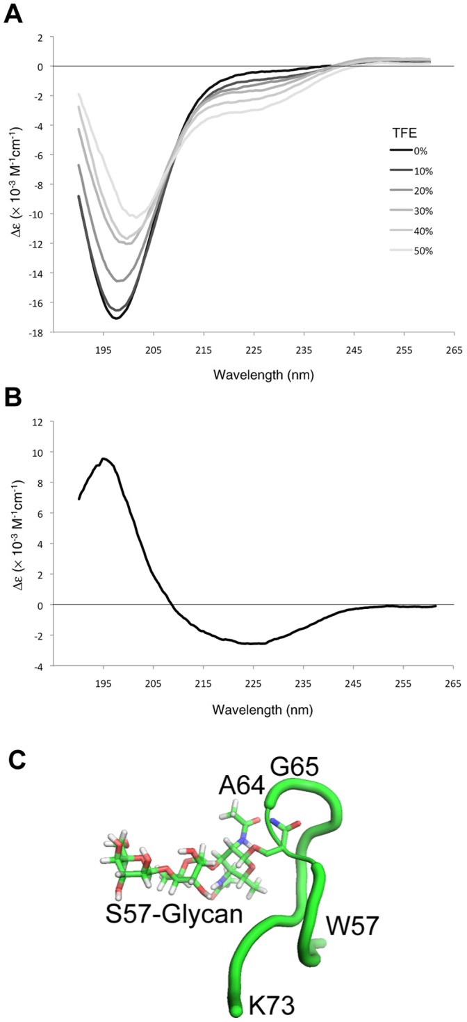
(A) CD Spectrum of AniA C-terminal peptide (NGAAPAASAPAASAPAASASEKSVY). Peptide was analysed in 50 mM KH2PO4 with 0–50% of TFE. (B) The difference in CD spectra between the peptide in 50% TFE and no TFE. (C) Modelling of N. meningitidis PilE glycosylation site structure. Peptide corresponding to the glycosylated region of C311 PilE (57WPGNNTS(Gal(β1–4)Gal(α1–3)2,4-diacetimido-2,4,6-trideoxyhexose)AGVASSSTIK73) constrained as in the structure of N. gonorrhoeae PilE was modelled.