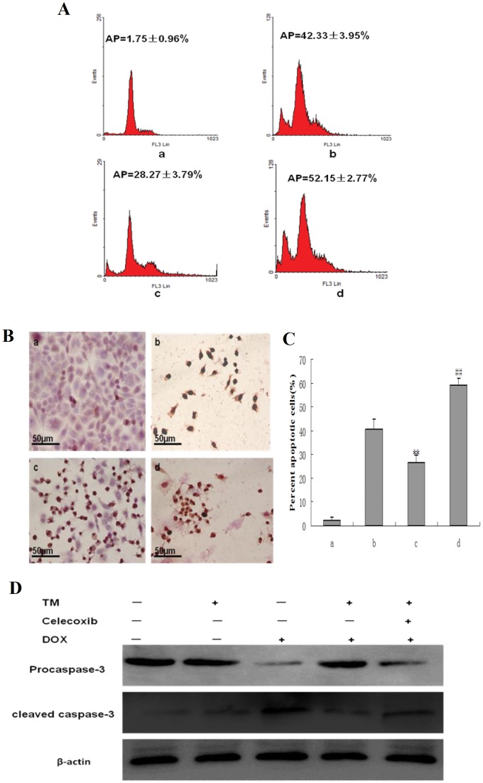
Figure 6. Effect of co-pretreatment with tunicamycin and celecoxib on apoptosis induced by doxorubicin in HepG2 cells.
(A) HepG2 cells were pretreated with 3 µmol/L tunicamycin for 8 hr, either in the absence or the presence of celecoxib (50 µmol/L) and then exposed to doxorubicin (2.5 mg/L) for 24 hr. Apoptosis was analyzed as the sub-G1 fraction by fluorescence-activated cell sorting (FACS). a: Untreated HepG2 cells; b: HepG2 cells treated with doxorubicin alone; c: HepG2 cells pretreated with tunicamycin, and then exposed to doxorubicin for 24 hr d:HepG2 cells co-pretreated with tunicamycin and celecoxib, and then exposed to doxorubicin for 24 hr. (B) and(C) Cell morphology and percentage of apoptotic cells was examined by TUNEL staining. a: Untreated HepG2 cells; b: HepG2 cells treated with doxorubicin alone; c: HepG2 cells pretreated with tunicamycin, and then exposed to doxorubicin for 24 hr d: HepG2 cells co-pretreated with tunicamycin and celecoxib, and then exposed to doxorubicin for 24 hr. Data are presented as mean ± SD for the three independent experiments. (**P<0.01 compared with HepG2 cells treated with doxorubicin alone, ##P<0.01 compared with HepG2 cells pretreated with tunicamycin, and then exposed to doxorubicin for 24 hr). (D) Cleaved caspase-3 as an apoptotic marker were measured by western blot using specific anti- caspase-3 antibody. β-actin in the same HepG2 cells extract was used as an internal reference.

