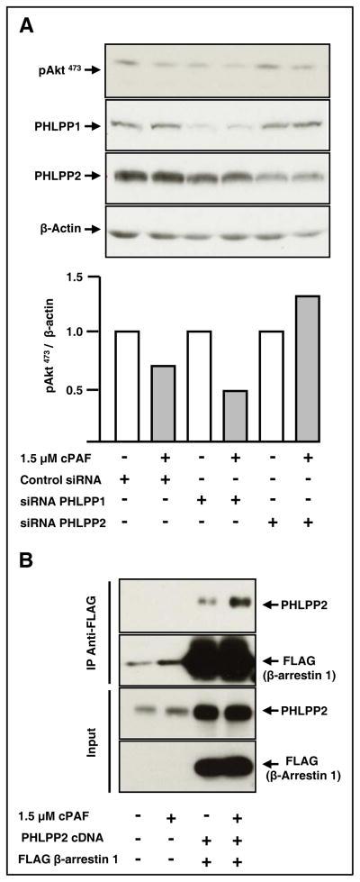
Figure 5. The Akt phosphatase PHLPP2 forms a complex with β-arrestin 1 and mediates cPAF-induced de-phosphorylation of pAkt473.
A. HT-29 cells were transfected with control oligos, or with siRNAs for PHLPP1 or PHLPP2, as described in Materials and Methods. After 24 hours at 37°C, the cells were starved overnight in serum-free media. We then stimulated the cells with cPAF for 45 min at 37°C. Cellular proteins were harvested, and then subjected to electrophoresis and immunoblot analyses.
B. HT-29 cells were transfected with cDNAs encoding PHLPP2 and FLAG-tagged β-arrestin 1. After 24 h at 37°C, we starved the cells overnight, stimulated them with cPAF for 30 min at 37°C, and then incubated cell lysates with anti-FLAG beads overnight at 4°C. We assessed PHLPP2 and FLAG-β-arrestin 1 levels in total lysates to determine transfection efficiency. We determined the extent of cPAF-induced association between PHLPP2 and β-arrestin 1 by subjecting the proteins associated with FLAG beads to electrophoresis and immunoblotting, using antibodies against PHLPP2 and β-arrestin 1.

