
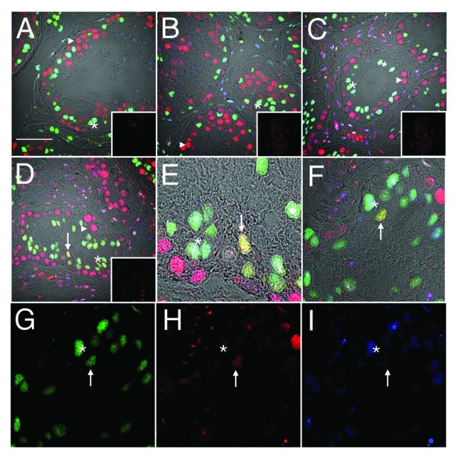
Figure 2. Downregulation of androgen receptor immunoreactivity in PCNA-positive Sertoli cells. Confocal immunofluorescence of human testis sections from normal (A) or gonadotropin-suppressed men (B, 2 wk; C–I, 12 wk). Sections were probed for GATA-4 [green, Sertoli cells within the seminiferous epithelium, asterisks), androgen receptor (blue, Sertoli (asterisks), Leydig and peritubular cells], and PCNA (red, labeling predominantly germ cells, triangles). Colocalization of GATA-4 and PCNA in Sertoli cells indicated by yellow (arrows). G–I are individual channels of the merged image in F, highlighting a GATA4 and PCNA-positive, AR-negative Sertoli cell (arrow) and a GATA4 and AR-positive, PCNA-negative Sertoli cell (asterisk). Inserts are controls where primary antibody was substituted for non-specific antibody of the same isotype. (Bar = 50 µm).