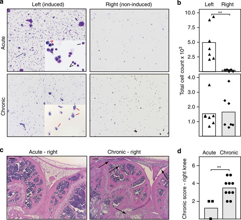
Figure 6. Spreading of arthritis to non-induced joints in chronic ACIA.
(a) Cytospins with May-Gruenwald-Giemsa staining of the synovial fluid of the left (induced) or right (non-induced) knee joint in the acute (4 days after induction) and chronic (5–8 weeks after induction) inflammation phase. Magnification X50 and X400. (b) Quantification of cells in the SF in the acute (top) and chronic phase (bottom). In the chronic phase a comparable number of cells is found in the synovial fluid of the induced and non-induced knee joints; the inflammation spreads to other joints. (c) HE staining of paraffin sections of the non-induced right knee joints in the acute (left) or chronic (right) phase of arthritis. Arrows indicate hallmarks of chronic inflammation: cell infiltrations, the hyperplastic synovial membrane and fibrosis. Black bars, 200 μm. (d) Scoring of histology sections for chronic inflammation in the non-induced (right) knee joint of BALB/c mice in the acute phase (day 4, squares) and chronic phase 5–8 weeks after induction of arthritis (circles). Scoring statistics: Mann–Whitney-U-test (n.s.: not significant, *P<0.05, **P<0.01, ***P<0.001).

