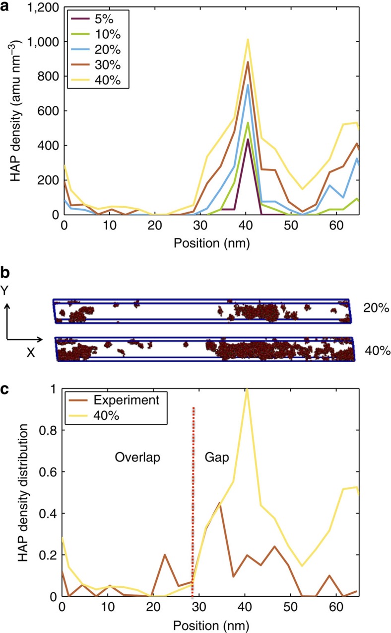
Figure 2. Mineral distribution in the collagen microfibril at different mineralization stages.
(a) Distribution of HAP along the collagen fibril axis. The data shows that the maximum amount of HAP is found in the gap region (between 30 and 50 nm). (b) Spatial distribution of HAP in the unit cell for 20 and 40% mineral density. (c) HAP density distribution along the fibril axis for the 40% case normalized (same data as depicted in panel a) compared with experimental data26. The comparison confirms that maximum deposition is found in the gap region.

