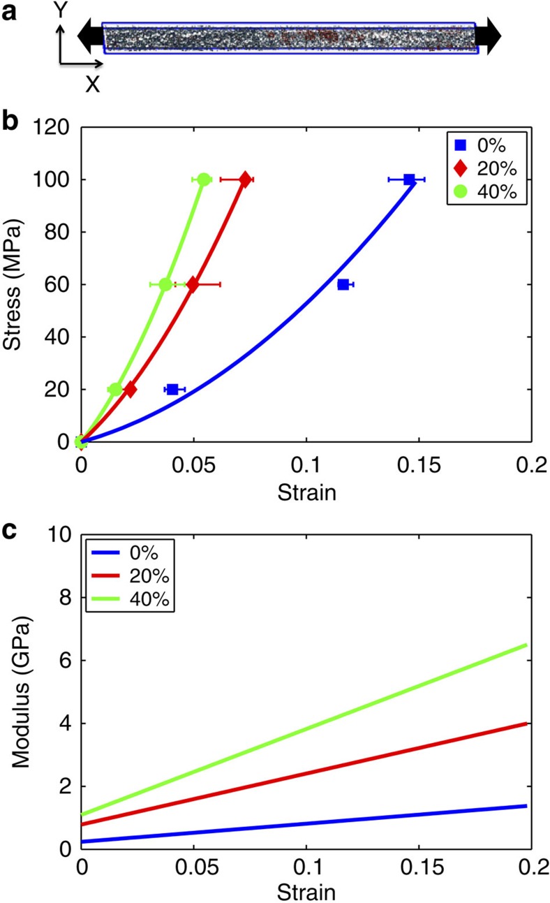
Figure 3. Mechanical properties of collagen fibrils at different mineralization stages.
(a) Fibril unit cell with mineral content used to perform tensile test by measuring stress versus strain. (b) Stress–strain plots for non-mineralized collagen fibril (0%), 20% mineral density and 40% mineral-density cases. (c) Modulus versus strain for 0, 20 and 40% mineral density showing an increase in modulus as the mineral content increases. The error bars in b are computed from the maximum and minimum values of the periodic box length along the x direction at equilibrium.

