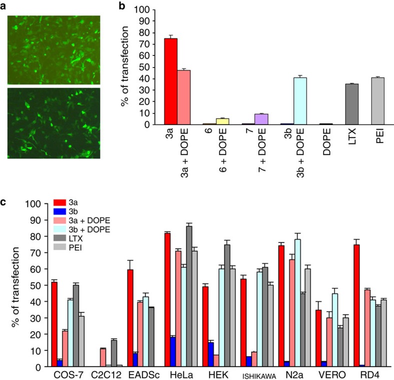
Figure 5. Cell transfection experiments.
(a) Images by fluorescence microscopy of human Rhabdomyosarcoma cells transfected (in green) upon treatment (at 48 h) with EGFP-C1 plasmid 1 nM formulated with (top) 10 μM calixarene 3a and (bottom) LTX. In histogram b, transfection efficiency (at 48 h) to Rhabdomyosarcoma cells of argininocalix[4]arenes 3a and 6 compared with the non-macrocyclic model 7 (20 μM), the lysinocalixarene 3b, DOPE (1,2-dioleoyl-sn-glycero-3-phosphoethanolamine), LTX and PEI (polyethyleneimine). Error bars denote s.d. (n>3). In histogram c, transfection efficiency to other cell lines of argininocalixarene 3a (red bar) compared with argininocalixarene 3a with DOPE (pale red bar), lysinocalixarene 3b (blue bar), lysinocalixarene 3b with DOPE (light blue bar), LTX (grey bar) and PEI (light grey bar). Calixarene derivatives and DOPE were used at 10 and 20 μM, respectively. Error bars denote s.d. (n>3).

