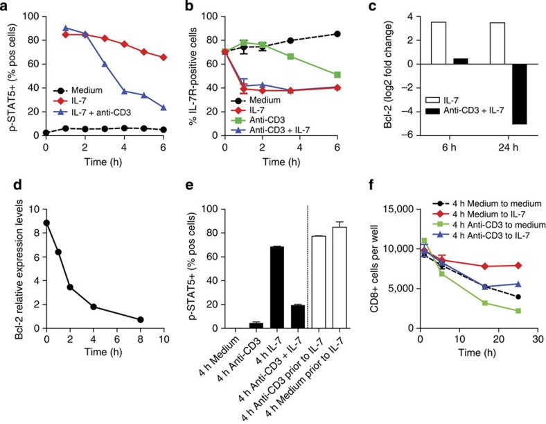
Figure 2. TCR ligation inhibits Bcl-2 and phosporylation of STAT5.
(a) Time course of percentage of cells positive for phospho-STAT-5 of CD4+ T cells from C57BL/6 mice cultured in medium alone, with 10 ng ml−1 IL-7 or stimulated with plate-bound anti-CD3 with 10 ng ml−1 IL-7 measured by flow cytometry. (b) Expression of IL-7Rα on CD8+ T cells from C57BL/6 mice cultured in medium or stimulated with plate-bound anti-CD3 with or without 1 ng ml−1 IL-7. IL-7Rα levels were measured by flow cytometry at indicated times and expressed as percentage IL-7Rα-positive cells. (c) Relative expression levels of bcl-2 mRNA in CD4+ T cells from C57BL/6 mice cultured in 1 ng ml−1 IL-7 alone or stimulated with plate-bound anti-CD3+1 ng ml−1 IL-7 measured by quantitative PCR (qPCR). Data are expressed as log2 of the fold change: bcl-2 mRNA expression relative to bcl-2 levels in freshly isolated cells. (d) Relative expression levels of bcl-2 mRNA of CD4+ from C57BL/6 mice T cells stimulated with anti-CD3 after overnight culture in IL-7 measured by qPCR. Data are expressed as expression relative to freshly isolated cells. (e) Loss of STAT5 phosphorylation is reversible after removal from antigen stimulus. Phosphorylation of STAT5 was measured by flow cytometry in CD8+ T cells unstimulated or stimulated with plate-bound anti-CD3 before exposure to IL-7. CD8+ T cells from C57BL/6 mice were cultured either continuously for 4 h in medium or on CD3 antibody-coated plates with or without 1 ng ml−1 IL-7 (filled columns) or were cultured in medium or on CD3 antibody-coated plates for 4 h without IL-7 before being removed from stimulus, and then exposed to 1 ng ml−1 IL-7 for 15 min (open columns). (f) Removal from TCR stimulation does not fully restore IL-7-mediated survival. CD8+ T cells C57BL/6 mice were cultured in medium or on anti-CD3-coated plates for 4 h before being removed from stimulus (indicated as 0 h), and cultured in new medium with or without 1 ng ml−1 IL-7 for up to 24 h. Data represent viable cells per well. (b, e, f) Data are presented as means±s.e.m. of triplicate cultures.

