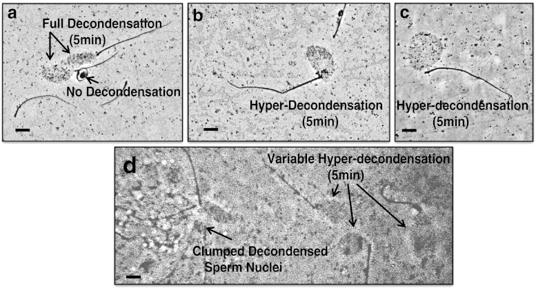
Fig. 2.
(Panels a-d): Microscopic images of a patient’s sperm with a positive (abnormal) SDAD Test score = 124.7 % of the negative control with fully decondensed sperm nuclei after a 5 min incubation in egg extract. All images are at the same magnification: Bar = 15 μm. Panel a: Microscopic images of 2 fully decondensed sperm nuclei by a phase dark sperm with no decondensation, and the sperm’s tail wrapped around the sperm head. Panels b and c: Typical images of hyper-decondensed sperm nuclei. Panel d: Clumped decondensed sperm nuclei, and variable hyper-decondensed sperm nuclei, all in the same field of view

