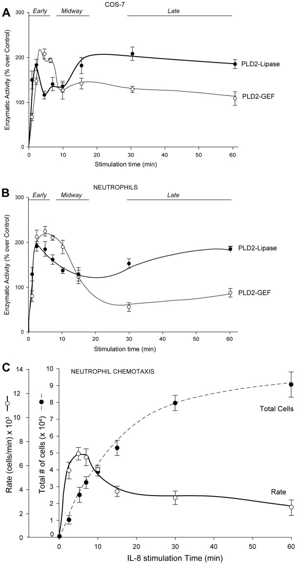
Fig. 6.
PLD2 lipase and GEF cellular activities as a function of time. Effect of EGF (A) or IL-8 (B) stimulation on increasing time of the lipase and GEF activities of PLD2 overexpressed in COS-7 cells (A) and primary neutrophils (B). PLD2's lipase and GEF activities peak at early time following EGF stimulation, whereas at later times lipase activity continues to increase but GEF declines. (C) Chemotaxis of primary neutrophils. Peripheral blood neutrophils migrated towards IL-8 in a time-dependent manner. Cells were incubated in transwell permeable supports in multi-well plates over a period of 60 minutes. The dashed line represents accumulation of neutrophils to the bottom well where the chemoattractant was added. The solid line represents the ‘rate’ of migration (as number of cells migrated per unit of time). Values are means± s.e.m. of three independent experiments performed in duplicate.

