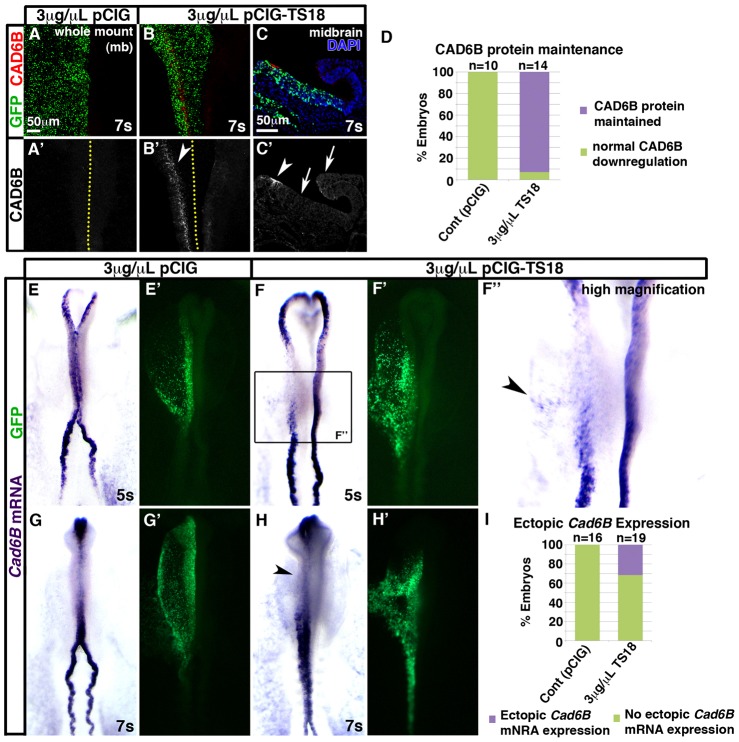
Fig. 6.
Tspan18 overexpression maintains Cad6B protein, while Cad6B mRNA downregulates on schedule. Embryos unilaterally electroporated with empty pCIG (A,E,G) or pCIG-TS18 (B,C,F,H) were immunostained for Cad6B protein at 7 somites (A,B, wholemount midbrain dorsal view; C, transverse midbrain section) or processed by whole mount in situ hybridization for Cad6B mRNA (E–H, dorsal view, anterior embryo half; green in A–C,E′–H′, construct targeting). (A–C) Cad6B protein (A–C, red; A′–C′) is downregulated normally in pCIG electroporated embryos (A′) but maintained in the midbrain dorsal neural tube of pCIG-TS18 electroporated embryos (white arrowheads in B′,C′). Ectopic Cad6B protein was not observed in the ventral neural tube or unelectroporated side of the embryo (white arrows in C′). Dotted lines in A′,B′ indicate embryo midline. (D) Bar graph represents the number of embryos at 7–10 somites exhibiting normal downregulation versus maintenance of Cad6B protein. (E–H) Although pCIG-TS18-electroporated embryos at 5 somites exhibit Cad6B mRNA expression in the neural folds that is dispersed (F) and sometimes ectopic (arrowhead in F″) compared with pCIG electroporated embryos (E), Cad6B mRNA is downregulated at 7 somites with the same temporal profile in pCIG (G) and TS18MO-electroporated embryos (H). (I) Bar graph represents the number of embryos exhibiting ectopic Cad6B mRNA expression. Scale bars: 50 µm (A,C).

