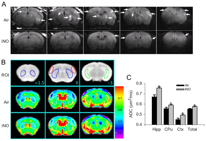
Fig. 3.

(A) Representative diffusion-weighted image (DWI) of mice brain 24 h after CA/CPR that breathed air (Air) or air supplemented with NO (iNO). White arrows indicate areas of hyperintense DWI. (B) Representative MR images showing three brain slices containing regions of interest [ROI]. Slice positions are identified in millimeters (+1.5, 0, or −3 mm) with respect to bregma in the coordinate space of the Allen Mouse Brain Atlas. Colored outlines indicate portions of ROI (blue, caudoputamen; red, lateral cortex; green, ventral lateral hippocampus) that intersect with these slice planes. Average ADC values of the slice plane for mice that breathed Air after CA/CPR [Air]. Average ADC values of the slice plane for mice that breathed NO after CA/CPR [iNO]. Color bar on the right side indicates color-code for ADC values (μm2/ms). (C) Average ADC values of each three-dimensional ROI (Hipp, ventral lateral hippocampus; CPu, caudoputamen; Cortex, lateral cortex; total, total brain) across all planes in mice that breathed air (Air, n = 6) or NO (iNO, n = 7) after CA/CPR. *P<0.05 vs. Air (Minamishima et al., 2011).
