Figure 1.
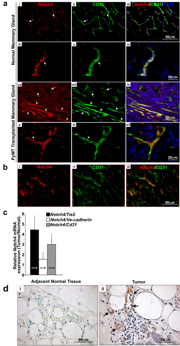
Notch4 expression is upregulated in the vasculature of mouse and human mammary tumors. (a) Notch4/CD31 immunostaining reveals an increase of Notch4 expression in the vasculature of mouse orthotopic MMTV-PyMT mammary tumors (vii-xii) compared to age and stage matched (virgin 8 weeks old) normal mouse mammary glands (i-vi). In panels vii-viii arrows indicate Notch4 low-expressing vessels; arrowheads indicate Notch4 positive vessels. Panels i to iii show low expression levels of Notch4 and CD31 in the vessels of normal mammary gland. Panels iv to vi show a detail of the staining of Notch4 and CD31 in the vessels of normal mammary gland. Panels vii-xii show Notch4 and CD31 staining in tumors without adjacent normal tissues in these images. Panels vii and ix show higher expression of Notch4 in vessels of the tumors than those in the normal gland (as compared to panels i and iii). Panels x (Notch4 staining), xi (CD31 staining) and xii (merged) show higher magnification of tumor vessels with Notch4 expression. (b) Notch4 staining in the tumor is mainly restricted to the vasculature. Orthotropic MMTV-PyMT mammary tumors at 3 weeks post transplant were immunostained for (i) Notch4, and (ii) CD31. Panel iii shows the merge of Notch4 and CD31. (c) Notch4 mRNA expression is increased in orthotopic tumors derived from transplanted MMTV-PyMT cells compared to normal mammary tissue as measured by quantitative RT-PCR. Data is represented as fold change of Notch4 expression in tumor over normal mammary glands, normalized to the expression of Tie2, VE-cadherin and CD31. Samples were analyzed in duplicate and error bars represent standard error of mean (s.e.m). (d) Notch4 immunostaining (brown stain) is increased in the vasculature of human infiltrating ductal carcinoma tissue (ii) compared to adjacent normal tissue (i). Arrows indicate Notch4 staining in the endothelium.

