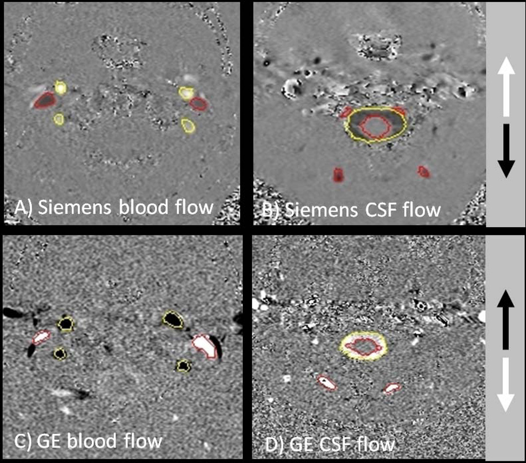
Figure 2.
Example of cine phase contrast image of blood flow and CSF flow taken on a Siemens and a GE system respectively. Note the difference of the color-scheme, where cranial inflow is encoded in white and cranial outflow is encoded in black on the Siemens system. The direction of the flow encoding is opposite on GE systems.

