Figure 3.

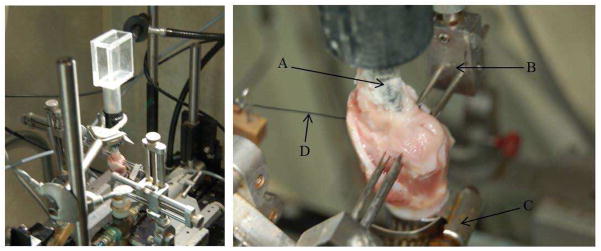
Left: Digital image of the experimental setup. Right: An enlarged view of the excised larynx setup highlighting A) epilaryngeal insert, B) three-pronged adduction device, C) tracheal hose clamp and D) the elongation suture.

Left: Digital image of the experimental setup. Right: An enlarged view of the excised larynx setup highlighting A) epilaryngeal insert, B) three-pronged adduction device, C) tracheal hose clamp and D) the elongation suture.