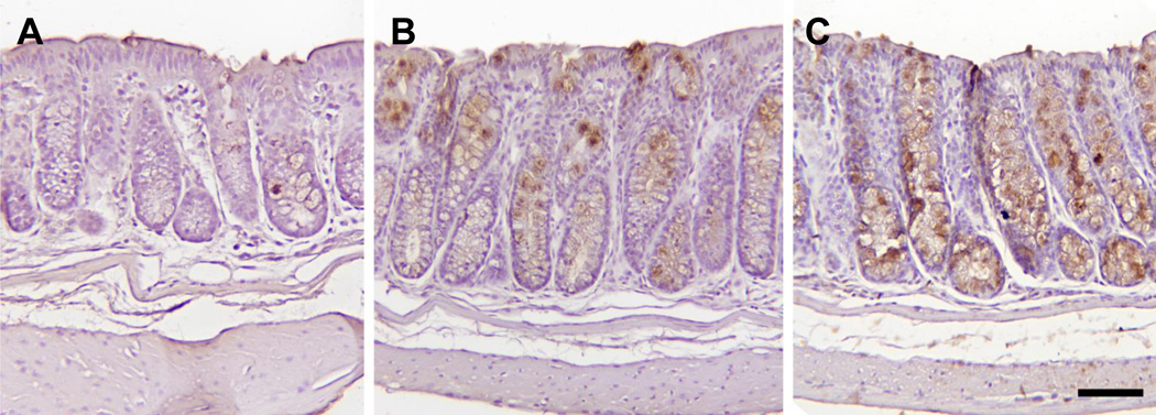
Fig. 4.
Representative images of MUC2 immunohistochemical staining of colon of A/J mice. The bar is equal to 75 micrometers. Immunohistochemical analysis was done as described in the Material and methods section. Microscopic photograph of colon tissue from (A) vehicle-treated mice, (B) 30 mg/kg MCC-555-treated mice, and (C) 60 mg/kg MCC-555-treated mice.

