
Figure 1. Wounds of adult zebrafish undergo rapid re-epithelialization.
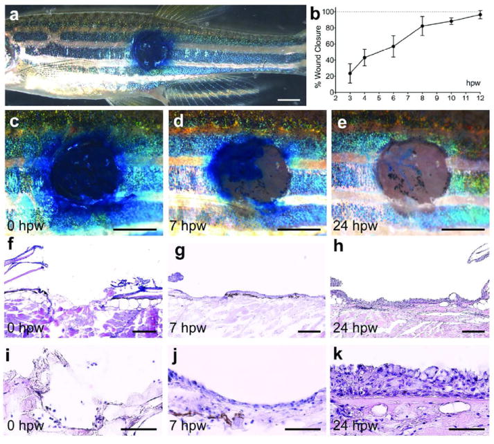
(a) Overview of the left flank of an adult zebrafish with an approximately 2 mm circular wound stained with methylene blue. (b) Graphical illustration of time course of wound closure (see Material and Methods); mean values of closure and standard deviations were determined for at least 6 individuals per time point using Excel software. (c-e) Magnified superficial views of the wound. At 0 hpw (a, c), methylene blue penetrates the entire wound, while the epidermal barrier has been partly recovered by 7 hpw (d), and fully recovered by 24 hpw (e). (f-k) H&E staining of longitudinal sections through the wound reveals the removal of epidermis, dermis and scales via the applied wounding protocol (f and I; n=4). At 7 hpw, a thin neo-epidermis is observed on the surface of the wound (g and j; n=6). At 24 hpw, the recovered epidermis appears re-stratified (h and k; n=10). Scale bars: a,c-e = 1 mm; f-h = 200 μm; i-k = 50 μm.