
Figure 2. Adult zebrafish exhibit a strong inflammatory response and granulation tissue formation.
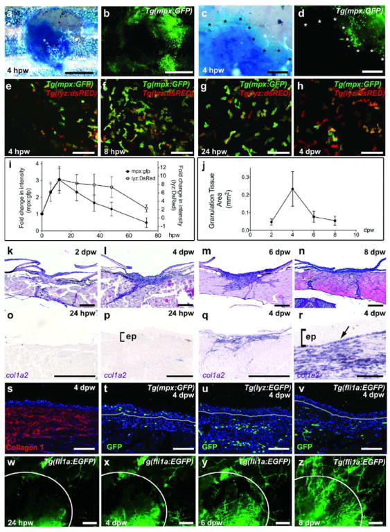
(a-d) Brightfield (a,c) and fluorescent (b,d) images of a Tg(mpx:GFP) fish at 4 hpw; GFP-positive neutrophils are present in re-epithelialized (methylene-blue excluding) regions of the wound (edges marked by asterisks in c and d; n=6/6). (e-h) Live images of the wound centre of Tg(mpx:GFP)i114/Tg(lyz:DsRED2)117 double transgenic fish reveal inflammatory cells, with a progressive relative increase of macrophages (h). (i,j) Graphical illustrations of time course of wound inflammation (i) and granulation tissue formation (j); mean values and standard deviations of relative fluorescent intensities (i) or granulation tissue areas (j) were determined for at least 6 individuals per time point using Excel software. (k-n) H&E staining reveals formation of granulation tissue beneath the wound from 2-6 dpw, which then regresses (8 dpw, n). (o-r) col1a2 expression beneath the wound is sparse at 24 hpw (o,p; n=4/4), but very prominent at 4 dpw (q,r; n=6/6). In addition to dermal fibroblasts, col1a2 is expressed by basal keratinocytes of the neo-epidermis (indicated by arrow in r). (s-v) Immunofluorescence analysis at 4 dpw reveals Collagen 1 deposition (s; n=4/4), leukocytes (t,u; n=4/4) and blood vessels (v; n=4/4) within the granulation tissue. mpx-positive neutrophils are also present in the neo-epidermis (t; n=4/4). (w-z) Superficial views of a Tg(fli1a:EGFP) fish shows progressive wound vascularization from 24 hpw to 8 dpw (n=4/4). Scale bars: a,b,o,q = 1mm; c,d, w-z = 250 μm; e-h = 50 μm; k-n = 500 μm; p,r,s-v = 100 μm. Abbreviation: ep, epidermis.