
Figure 3. Adult zebrafish exhibit minimal scar formation.
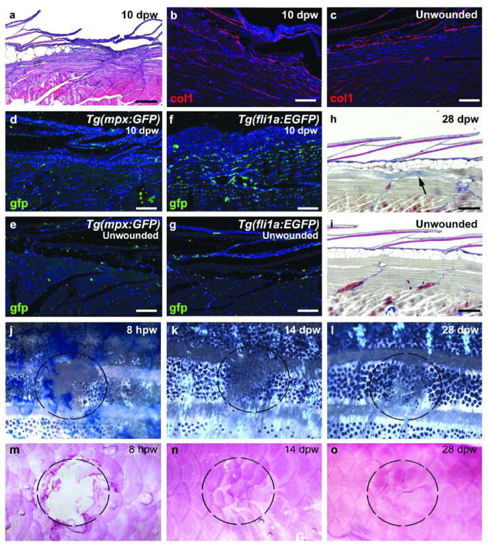
(a) H&E staining at 10 dpw reveals minimal remaining granulation tissue and newly forming scales. (b,c) At 10 dpw, minimal Collagen1 deposition is observed beneath the regenerating scales (b), similar to an unwounded region (c) (n=6/6). (d-g) Tg(mpx:GFP) (d,e; n=4/4) and Tg(fli1a:EGFP) (f,g; n=4/4) transgenic fish at 10 dpw display normal number of leukocytes (d) and reduced numbers of blood vessels (f) at the regenerating wound site when compared to an unwounded region (e and g). (h,i) AFOG staining at 28 dpw indicates the complete recovery of epidermis, dermis, scales and adipocytes with rarely occurring collagen deposits within the muscle layer beneath (h; n=4/4), compared to unwounded fish (i). (j-o) Superficial views of wounded fish demonstrate the almost complete recovery of stripe pattern (j-l) and scales (m-o; alizarin red) by 28 dpw (n=6/6). Dashed circles mark the position of the wound. Scale bars: a,h,i = 500 μm; b-g = 200 μm.