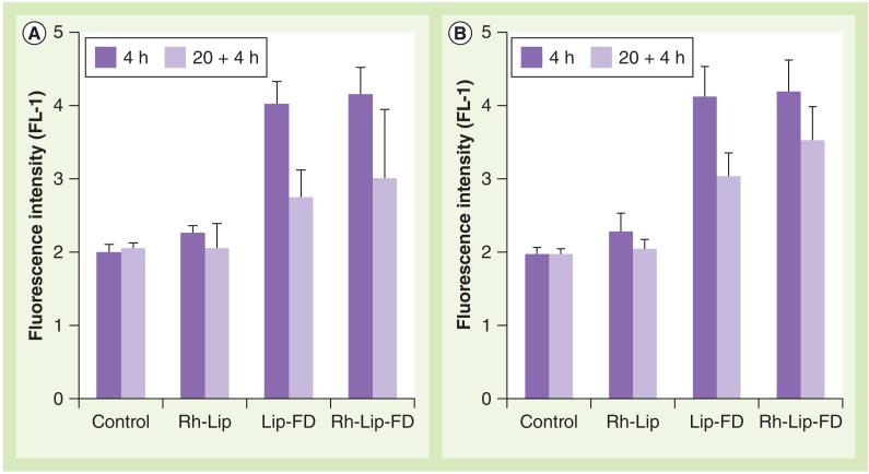
Figure 2. Interaction of liposomes with monocyte-derived macrophages.
Macrophages had (A) active or (B) inhibited lysosomal glucocerebrosidase. Activity of glucocerebrosidase in monocyte-derived macrophages was inhibited by treatment of the cells with conduritol B epoxide (200 μM). The cells were treated with equal amounts of plain Lip-FD, Rh-Lip or Rh-Lip-FD for 4 h (dark bars) followed by washing and additional incubation for 20 h in liposome-free medium (pale bars). The cells were then treated with 5-(pentafluorobenzoylamino)fluorescein diglucoside and analyzed by flow cytometry. The data represent the mean of three different experiments ± standard deviation.
Lip-FD: Fluorescein isothiocyanate-dextran-loaded plain liposomes;
Rh-Lip: Octadecyl-rhodamine B-modified liposomes; Rh-Lip-FD: Fluorescein isothiocyanate-dextran-loaded octadecyl-rhodamine B-modified liposomes.

