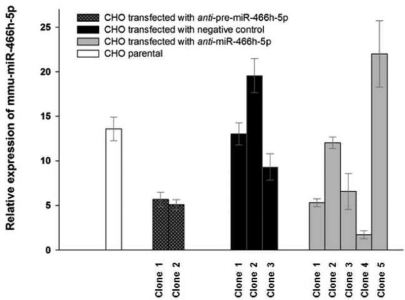
Figure 1.
Relative expression of mmu-miR-466h-5p in single CHO cell clones after PBA treatment. Single colonies of parental CHO and CHO transfected with negative control, anti-pre-miR-466h and anti-miR-466h-5p shRNAs were treated with PBA to compare activation of mmu-miR-466h-5p TaqMan qRT-PCR assays were used for analysis with snoRNA202 used as control for 2-ΔΔCt analysis. The levels of mmu-miR-466h-5p in each clone after PBA treatment were related to its levels in the same clone treated with solvent.

